What is fetal CTP, and how should it be normal during pregnancy?
How a baby grows and whether everything is fine with him, the doctor and spouses who are preparing to become parents will be helped to learn ultrasound diagnostics. At the very beginning, when the size of the baby is still very microscopic, the main criterion for the well-being of the baby is the so-called KTR. About what it is, how it can be, and what to do if there are deviations, we will tell in this material.
What it is
The abbreviation "KTR" is not an analysis or a research method, but the name of one of the sizes, which are determined on an ultrasound scan by a somnologist. Abbreviation is a complex term - coccyx parietal size. This concept means the distance from the highest point of the crown of the embryo and fetus to the lowest point of its tailbone in a position in which the body of the baby is completely unbent.
KTR is not a growth or a total length, as some people think. It's just cut from the head to the extreme point of the future spinebut for now - the neural tube. This parameter is measured from the earliest dates of gestation and up to 14 weeks.
After this, the baby becomes too large for the ultrasound sensor to cover such a distance at a time, and individual dimensions of the baby’s body parts come first, according to which the doctor judges the proportions, growth rates and development of the fetus.
KTR begin to measure almost immediately after the fact of pregnancy becomes apparent. To find out if there is a pregnancy at all, a woman with an ultrasound scan can take about 5 weeks, that is, 21 days after ovulation or about a week after the start of the delay of the next menstruation.
Kopchiko-parietal size is possible to measure after about a week, on the sixth obstetric week, that is, about a month from conception.
Growth rates of KTR inform the doctor about how the baby grows. In the early stages - this is the only thing that can talk about the well-being or unhappiness of pregnancy. The values of the coccyx-parietal size are supplemented with data on the viability of the crumbs, because from about 5 weeks on the ultrasound you can see the heartbeat.
The tiny heart is not yet fully formed, but the characteristic ripple is not hidden from the experienced gaze of a good specialist and a modern ultrasound scanner equipped with a sensor with high image detail.
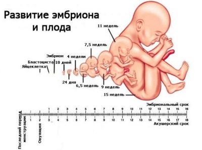
All babies in the first weeks of pregnancy grow at about the same rate. A little later, when the genetic characteristics of the child begin to appear, the ultrasound data from different pregnant women will have significant differences. With two pregnancies occurring on the same day and even an hour, the children will differ in size and body proportions, because they have different parents in terms of complexion, different heredity, different conditions of intrauterine growth.
In the short term, all embryos are almost the same, and they develop not because of hereditary peculiarities, but according to the laws of nature. That is why KTR is considered one of the most accurate methods for determining the duration of pregnancy. Accuracy at the same time - until every day after conception.
KTR does not depend on race, color, nationality of parents, on ecology and geographic latitude. The only thing that is theoretically capable of influencing the coccyx parietal size is features of the health of both the expectant mother and the baby himselfAfter all, even for a short time, literally from the first minutes after conception, all the information about how the baby will be healthy is incorporated in its genetic code - the karyotype.
How is it measured?
The only way to measure CTE is sagittal scanning. If the baby is moving too intensively, the doctor waits for the moment when he will fully bend to make a freeze-frame.
The sagittal plane visually divides the small body in half. The length that the doctor lays from crown to the coccyx is measured by a special program embedded in the scanner. The resulting value correlates with existing tables that indicate normal CTE values depending on the duration of gestation. Accuracy of the study - plus or minus 3-4 days.
In this way, this size allows you to set the exact duration of pregnancy in the case when it is necessary. Such a need may occur in women suffering from irregular menstrual cycles, not remembering the exact date of the beginning of the last menstrual period. This date is considered the starting point of obstetric gestational age.
The first day of the last menstruation is considered the first day of pregnancy. That is, obstetric period differs from the actual by almost 2 weeks or more.
The kopchiko-parietal size of the fetus will allow you to establish the exact time in case the doctor suspects embryo development, because it is possible that ovulation occurred later than the woman thinks, or implantation occurred somewhat later than stipulated by the average statistical period. Then the actual age of the fetus will be less, which is what the KTR will determine on the ultrasound scan.
KTR norms
As already mentioned, embryos grow at approximately the same rate if the pregnancy develops safely. The existing tables are considered to be a fairly reliable way to assess growth and clarify the duration of pregnancy. Therefore, in relation to the coccyx parietal size in the early stages, no possible fluctuations of the parameter being determined are indicated in a smaller direction.
Values are quite accurate. But it is only on the smallest terms. As the baby grows, the permissible errors in KTR appear - the upper and lower thresholds of the norm.
Table of normal values of CTE and compliance with the term of gestation:
Gestational age (week + day) | The lowest value of KTR, mm | The average value of the CTE, mm | Maximum value of CTE, mm |
5+0 | Not determined | 1,2 | 4,3 |
5+1 | Not determined | 1,4 | 4,6 |
5+2 | Not determined | 1,7 | 4,9 |
5+3 | Not determined | 2,0 | 5,3 |
5+4 | Not determined | 2,3 | 5,7 |
5+5 | Not determined | 2,7 | 6,1 |
5+6 | Not determined | 3,1 | 6,6 |
6+0 | Not determined | 3,5 | 7,1 |
6+1 | 0,4 | 4,0 | 7,7 |
6+2 | 0,8 | 4,6 | 8,3 |
6+3 | 1,3 | 5,1 | 8,9 |
6+4 | 1,9 | 5,8 | 9,6 |
6+5 | 2,5 | 6,4 | 10,4 |
6+6 | 3,1 | 7,1 | 11,1 |
7+0 | 3,8 | 7,9 | 11,9 |
7+1 | 4,5 | 8,7 | 12,8 |
7+2 | 5,3 | 9,5 | 13,7 |
7+3 | 6,1 | 10,4 | 14,7 |
7+4 | 6,9 | 11,3 | 15,6 |
7+5 | 7,8 | 12,2 | 16,6 |
7+6 | 8,7 | 13,2 | 17,7 |
8+0 | 9,7 | 14,2 | 18,8 |
8+1 | 10,7 | 15,3 | 20,0 |
8+2 | 11,7 | 16,4 | 21,1 |
8+3 | 12,8 | 17,5 | 22,3 |
8+4 | 13,9 | 18,6 | 23,5 |
8+5 | 15,0 | 19,9 | 24,7 |
8+6 | 16,2 | 21,1 | 26,1 |
9+0 | 16,3 | 22,0 | 27,0 |
9+1 | 17,0 | 23,0 | 29,1 |
9+2 | 18,1 | 24,2 | 30,0 |
9+3 | 19,0 | 25,0 | 31,0 |
9+4 | 20,2 | 26,1 | 32,0 |
9+5 | 21,0 | 27,0 | 34,1 |
9+6 | 22,1 | 29,0 | 36,0 |
10+0 | 24,2 | 31,1 | 38,0 |
10+1 | 25,3 | 33,1 | 41,0 |
10+2 | 26,0 | 34,0 | 42,0 |
10+3 | 27,1 | 35,0 | 43,2 |
10+4 | 29,0 | 37,1 | 45,0 |
10+5 | 31,0 | 39,3 | 47,2 |
10+6 | 33,0 | 41,1 | 49,0 |
11+0 | 34,0 | 42,2 | 50,1 |
11+1 | 35,1 | 43,0 | 51,0 |
11+2 | 36,0 | 44,2 | 52,1 |
11+3 | 37,0 | 45,3 | 54,1 |
11+4 | 38,1 | 47,0 | 56,0 |
11+5 | 39,0 | 48,2 | 57,1 |
11+6 | 40,1 | 49,0 | 58,3 |
12+0 | 42,0 | 51,3 | 59,0 |
12+1 | 44,0 | 53,0 | 62,0 |
12+2 | 45,1 | 55,0 | 65,1 |
12+3 | 47,0 | 57,1 | 67,1 |
12+4 | 49,1 | 59,3 | 69,2 |
12+5 | 50,0 | 61,2 | 72,0 |
12+6 | 51,0 | 62,0 | 73,4 |
13+0 | 52,1 | 63,0 | 75,0 |
13+1 | 53,1 | 65,2 | 77,0 |
13+2 | 54,0 | 66,0 | 78,2 |
13+3 | 56,0 | 68,1 | 80,0 |
13+4 | 58,0 | 70,0 | 82,1 |
13+5 | 59,2 | 72,0 | 85,2 |
13+6 | 61,0 | 74,0 | 87,2 |
14+0 | 63,0 | 76,0 | 89,0 |
In this way, establish the exact duration of pregnancy is not work. The values in the ultrasound protocol for a particular woman can be specified without the tenths, only integers. It all depends on how modern the device and the sensitive sensor were during the measurement.
Older machines define values in integers. The latest generation of scanners, which are equipped with modern perinatal centers and clinics, can determine the CTE with an accuracy of tenth.
You should not try to “fit” your values to average values up to every tenth. If according to the results of the ultrasound it is written that the fetal CTE = 1 mm, it can be both 1.1 and 1.5 mm. If it is indicated that КТР = 23,7, this corresponds to 9 weeks and 1 day from conception with an error of + 1 day. Similarly, CTE = 61 mm corresponds to a period of 12 weeks and 5 days, although in the table the tenths of different values are different.
Possible problems and their causes
During pregnancy, women become incredibly sensitive, especially to everything related to the development and health of the baby. If there are no special problems in the early stages, the woman does not complain about anything, then there is no need to go for an ultrasound before 10-13 weeks.At the time indicated, the first “meeting” of the mother with the baby usually takes place.
Naturally, it is the KTR of the fetus and all related issues and issues that becomes an important topic for discussion in women's forums and in the communities of parents. The most common of these should be considered in more detail.
KTR is less than normal
This situation is common, it is she who causes the greatest amount of suffering and worries for future mothers. The lag of the coccyx-parietal size from the norm can be both physiological and pathological. For example, at week 11, the kid on the size of KTR was barely close to week 10. Naturally, the pregnant woman begins to get upset and look for the causes of this phenomenon.
Not everything is so bad, experts say. After all, most often the reason lies not in the pathologies of the fetus and not in the diseases of the mother, and in the banal error that crept into the timing calculation of pregnancy. When a woman visits a gynecologist about the delay of menstruation, and the doctor confirms the fact of pregnancy, he is counting from the first day of the last menstrual period.
If the woman made a mistake with the date, then the deadline will be set incorrectly. If ovulation is late or implantation of the ovum into the uterus occurs later, then the dispenser will be less than the norm just for the period for which it was delayed.
Check or refute this version will help re-ultrasound, which is usually assigned a week and a half after the first ultrasound scan. In case of an error in calculating the term, the KTR will increase in proportion to the actual term, and further pregnancy management will be carried out according to the corrected data.
The cause of the lag of the coccyx-parietal size from the existing normative values may be the death of the fetus. If the baby for some reason has ceased to develop, and the miscarriage has not occurred, his KTR will be at the level at which he is at the time of his death.
If the term is still small, and it is difficult to reliably determine the fact of fetal heartbeat, Repeat ultrasound is desirable to spend in 5-6 daysto make sure that the baby is not growing.
In this case, obstetric curettage of the uterus or drug-induced abortion without surgical intervention (with a fetal period of less than 6 weeks) is indicated. Perhaps further treatment with antibiotics.
In obstetric curettage, embryo tissue samples are taken for genetic analysis in order to determine the cause of his death. Most often it lies in total chromosomal abnormalities, in which further development of the embryo was impossible.
A lag in KTR values of more than two weeks is a reason for a more detailed examination. If the baby is alive and growing, but its piggy-parietal size is significantly different from the norm, the reason may be hidden in the hormonal background of the mother.
Lack of such a hormone as progesterone, affects the development of the baby at the very beginning. If the blood test shows that the hormone is not enough, then the woman is prescribed hormone therapy. With drug support, the hormonal background is normalized, and the baby will quickly begin to recruit what he “missed”, KTR will become normal.
The reason for the reduction of the coccyx-parietal size may be the distress he is experiencing due to his mom's infectious or chronic diseases. Projections in this case depend on the specific disease or infection. If there is an opportunity to begin treatment, the doctor will prescribe it.
KTR is over normal
Kopchiko-parietal size may be more than the norm, and this also worries expectant mothers. Excess of this indicator is also characteristic of women who can not accurately remember the date of the last menstruation. An error in calculating obstetric terms can also occur in women with early ovulation.
Earlier, the release of the egg can occur due to a recent infectious disease, for example, influenza or ARVI, as well as in women who took hormonal preparations for the treatment of infertility.
Thus, the period may be longer than the doctor expects, and the size of the KTR will clearly indicate this. In order to confirm or deny the version, a woman will be asked to come back to the ultrasound doctor's office after about 10 days.
If the KTR will grow symmetrically to the date, and at the same time will consistently exceed the norm by days and weeks, further maintenance will be carried out taking into account these corrections. Deadlines recalculate, the expected date of birth will transfer to an earlier date.
If the KTR of the little ones is constantly at the limit of the upper norm or exceeds it, the doctors may suspect that the woman has a tendency to develop a large or even giant fetus.
Large in obstetrics is the baby, who is born with a weight of over 4 kilograms. A giant fetus is a child that weighs more than 5 kilograms at the time of birth.
Such women are not recommended to take vitamin complexes, eat a lot, expectant mothers should visit the doctor more often. The diagnosis “large fetus” is not officially set at the beginning of pregnancy. This happens much later, in the third trimester. But the suspicion of a large or giant baby may arise in the early stages.
It is not necessary to assume that the tendency to a large baby is the excess of KTR from the norm by 1-2 mm. A doctor can talk about trends when, for example, at week 13 of the KTR, with a norm of 63 mm and an upper threshold of 75 mm, there will be more than 80 mm.
Before assuming the hero, the mother will check the blood sugar level. Often women with diabetes have a predisposition to the birth of large children. Such a pregnancy will require special reference and attitude.
KTR is growing slowly
Sometimes a woman may face a particular problem - the slow growth of KTR. Such a phenomenon is said if the embryo shows signs of life (from 5 weeks - a heartbeat, from 8 - motor activity), but at the same time the growth of the crumbs is slowed down and over time more and more lags behind the existing norms. There are several reasons for this phenomenon, almost all of them are pathological.
Growth rates can slow because of genetic pathologies. While the period is too small, it is not possible to establish this reliably. The first screening, which takes place between 10 and 13 weeks inclusive, will be able to clarify the situation.
If the baby has congenital abnormalities, special markers in the blood of a pregnant woman on an ultrasound pattern will affect the establishment of a high risk for Down syndrome, Patau, Turner or other pathologies.
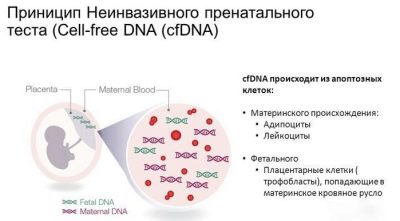
It is clear that waiting and tormenting a woman is absolutely unbearable. The only way to find out if the baby is healthy before screening is a non-invasive prenatal DNA test. It can be performed after 8 weeks of pregnancy in the medical genetic centers and genetic clinics.
For analysis, blood is taken from a vein, individual child red blood cells can be detected in it. Baby DNA is extracted from them, and the probability of a chromosomal abnormality is determined from the karyotype.
The analysis is very expensive - several tens of thousands of rubles, it is not available to everyone. This analysis is not included in the compulsory medical insurance package, so it will not work to do it for free.
The reason for the slow growth of the embryo can be a failure of the endometrium of the uterus, if not so long ago, the woman had an abortion. After the termination of pregnancy it is recommended to withstand a certain period before the next pregnancy, but in fact it does not always work and not at all.
If this happened, and in addition to the low growth rate of the CTE, the doctor states erosion of the cervix or inversion of the cervix in the direction of the vagina (ectropion), then the predictions are rather doubtful. The risk of miscarriage is very high.A woman needs hospitalization, in the hospital there are chances to save the baby and give him the opportunity to grow and develop according to the deadline.
Slow "gain" KTR is peculiar and Rh-conflict pregnancy. If a woman with negative Rhus has already had several genera or caesarean sections, and Rh-positive babies have appeared, then the sensitization of the maternal organism can be so strong that antibodies begin to grow literally from the first weeks of the next pregnancy.
In this case, the forecasts are also quite doubtful. Everything will depend on the specific situation, the level of antibodies in the mother's blood.
KTR is growing too fast
Such situations occur infrequently, but they can not be excluded. On average, up to 12 weeks the growth of the baby is increased by 1 mm per day, after this period the grit starts to grow faster. If the measurements were made at week 11, and then the ultrasound scan was repeated for some reason at week 14, then the growth may indeed look swift.
If growth increases above the norm and at an earlier date, then the main reasons that doctors will consider are metabolic disorders in the maternal organism, congenital abnormalities of the fetus and Rh-conflict. Maternal problems with hyperfunction of the thyroid gland can be accompanied by rapid growth of the fetus by week.
See how the fetal CTE is measured in the next video.
