Tetralo fallo in children
If a heart disease is detected in the crumbs, this cannot but cause feelings and anxiety in the parents. Especially if the diagnosis sounds incomprehensible and ominous, for example, Fallot's tetrad. What is the pathology behind this name and is it dangerous for the child?
What is it
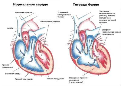
Fallo's notebook is one of the congenital cardiac pathologies, which is a combination of four abnormalities in the development of the heart. For the first time such a complex of defects was described by the French doctor Fallot, and therefore the disease bears his name. The tetrad includes such anomalies:
- The narrowing of the area through which blood leaves the right ventricle. It can be represented by stenosis of the valves, narrowing under the valves, as well as narrowing of the pulmonary trunk or stenosis of the pulmonary arteries.
- Pronounced defect of the septum separating the ventricles. As a rule, it is tall, that is, located close to the aorta. Because of such a defect, the right parts of the heart are connected to the left and the blood mixes.
- The displacement of the aorta in the right side, which is called dextraposition. Due to this altered position, the vessel may partially depart from the right ventricle.
- Thickening of the right ventricle, called hypertrophy. It develops as a result of difficulty in releasing blood from this chamber of the heart.

Fallot's tetrad is diagnosed in every tenth child with a congenital heart defect. This pathology takes about 50% of all defects with discharge of blood to the left.
The reasons
Fallot's tetrad is a congenital defect, since the anomalies observed with it are formed in utero. The appearance of this pathology is associated with a violation of the laying of the heart in the first 8 weeks of the development of the fetus, which can provoke:
- Infection, such as rubella or measles.
- Heredity.
- Ionizing radiation.
- Taking medicines, for example, sleeping pills or hormonal drugs.
- Alcohol consumption.
- Harmful working conditions.
- Drug use.
- Chromosomal disease.

Symptoms
The main clinical manifestation of Fallot's tetrad is cyanosis, due to which this pathology is referred to as "blue" heart defects. At the time of occurrence of such a symptom and its severity affects the degree of narrowing of the pulmonary artery. If cyanosis appeared in the first days of life, this defect is extremely serious. Most often, cyanosis develops slowly to the age of three months to a year, and in mild cases, its appearance is observed at the age of 6-10 years.
The skin tint in a child with Fallot's notebook may be different - either soft blue or dark blue or blue crimson. There is also an Acianotic form, in which the skin remains pale. Blueing is first observed on the lips, then on the mucous membranes and finger tips. Further, the child turns blue face, after which cyanosis spreads to the skin of all the limbs and torso. Hue is enhanced by the activity of the baby.
Another constant symptom of Fallot's tetrad is dyspnea. The child breathes deeply and arrhythmically, while the breathing rate practically does not increase. Dyspnea in a baby with such a pathology is noted at rest, and under any load, even the most minimal, it increases very sharply.
Children with such a defect gradually lag behind in physical development. They quickly enough appear changes of the fingers, which are called "watch glasses" (changing the shape of nails) and "drum sticks" (changing the shape of the phalanges).
For severe malformations in infants up to 2 years old, dyspnea and cyanosis attacks are diagnosed, the occurrence of which provokes severe brain hypoxia. During such attacks, children lose consciousness, and their risk of coma and death increases. The duration of the attack ranges from 10 seconds to several minutes. After the attack, the baby is sluggish and weak. Sometimes ischemia of the brain or hemiparesis develops.
Phases
In the clinical course of Fallot's tetrad, the following stages are distinguished:
- Phase 1 - lasts from birth to 6 months of age. Since the child feels satisfactory, this phase is called the state of relative well-being. Lag in development at this age is not observed.
- Phase 2 - lasts from 6 months to 2 years of age and is characterized by the appearance of attacks of dyspnea and cyanosis. In this phase, frequent cerebral complications and deaths are noted.
- Phase 3 - begins with 2 years of age. Attacks become less common and disappear due to the development of collaterals.
Diagnostics
Examination of a child suspected of Fallot's tetrad begins with an examination. The chest of such babies is often flattened, and the hump is absent. Listening to the heart of the baby, the doctor diagnoses a rough noise in the systole to the left of the sternum. Additional methods for detecting a defect are:
- Ultrasound of the heart. Anatomical defects that belong to the tetrad are determined.
- X-ray of the chest. The shadow of the heart in the picture resembles a shoe or felt boot.
- ECG. The axis of the heart deviates to the right, there are signs of an increase in the right side of the heart and conduction disorders.
- Sounding the heart. An increase in pressure in the cavity of the right ventricle, as well as low oxygen saturation of arterial blood is detected.
- Aortography Displacement of the vessel and the presence of collaterals is detected.
Treatment
In identifying Fallot's tetrad to a child, only surgical treatment is indicated, which is:
- Palliative surgery. It is performed at the first stage to children younger than three years old in order to relieve their condition and increase blood flow to the lungs. For this, anastomoses can be created or valve leaflets can be dissected.
- Radical operation. It is carried out 2-6 months after the first, connecting the baby to the artificial blood flow and cooling his body. During the intervention, stenosis in the right ventricle is eliminated and a patch is sutured to the interventricular septum.
In the postoperative period, attention is paid to strengthening the myocardium and reducing the load on the heart. The child is prescribed remedial gymnastics and a diet that allows him to recover quickly after surgical treatment.
Forecast
If Fallo's heavy tetrad is not treated, 25% of children with this pathology die before the age of one. The rest of the kids live on average 12 years and only about 5% of patients with this defect live to 40 years. The most common cause of death is brain damage due to the formation of a blood clot or abscess.
Surgical treatment at an early age is highly effective. Children after surgery are active and tolerate exercise. They must be observed by a cardiologist, and in any surgical and dental procedures, antibiotics are given to such children in order to prevent the appearance of endocarditis.
In the following video clip, Elena Malysheva will examine in more detail the topic of Fallo's tetrado disease in a child and the methods of treatment for this diagnosis.


